H1 SEO Custom Title
Dog & Cat Diagnostic Imaging
Kulshan Veterinary Hospital offers a wide range of diagnostic imaging options.
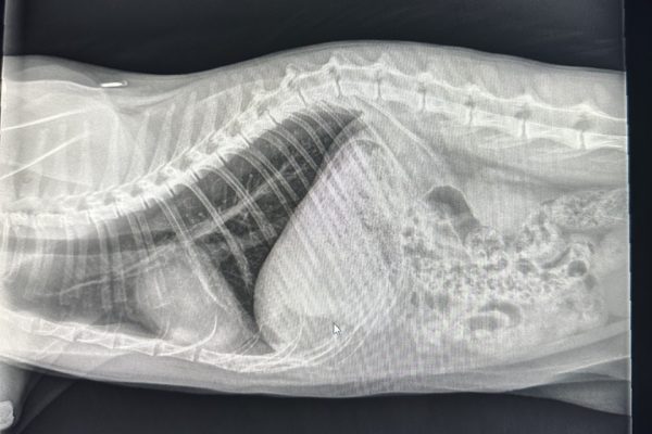
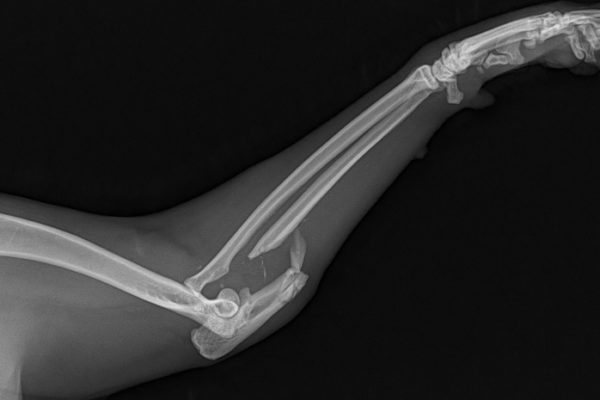
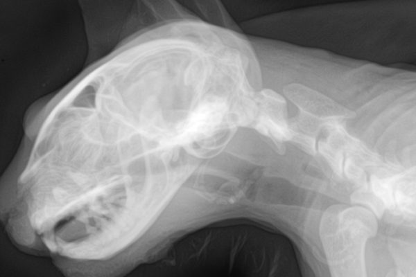
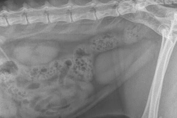
Radiography. We routinely take plain film radiographs of patients when indicated by their medical condition or history. This includes, but is not limited to, cases of possible fractures, foreign body ingestion, bladder stones and arthritis. Abdominal radiographs are frequently used just prior to pets giving birth to count the number of puppies or kittens an expectant mother is due to deliver. In more complicated case, contrast studies are done which involves using a special material that shows up on radiographs. It is used to enhance what is seen and assist in the diagnostic process. This technique is most commonly used in evaluating the GI tract or bladder.
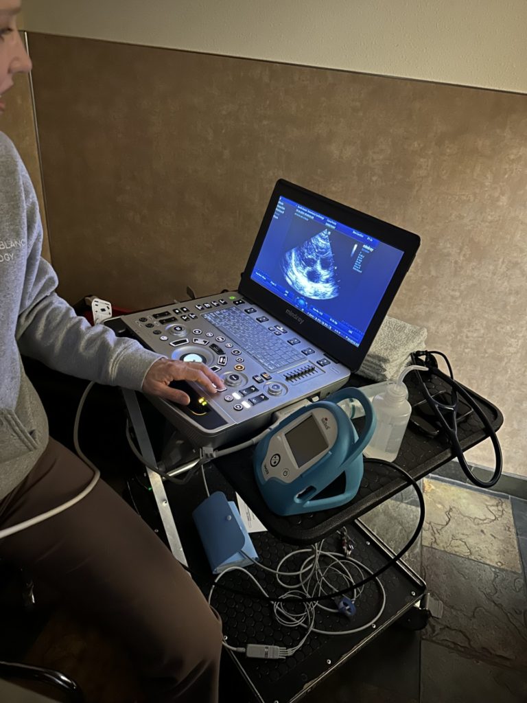
Ultrasound. Ultrasound uses sound waves to assist our doctors in evaluating their patients. It works best for looking at soft tissues such as the liver, kidneys, heart and bladder. It is also an exciting tool used for diagnosing and assessing pregnancy.
Endoscopy. Visualizing the inside of your pet is another diagnostic tool available at Kulshan Veterinary Hospital . Through the use of our state-of-the-art video endoscope, we can run a video camera down the esophagus into the stomach and proximal intestines to look for lesions, take biopsies and even retrieve some foreign materials.
Video Otoscopes. These small endoscopes are connected to a video camera unit used to examine the outer ear, ear cannal and tympanic membrane. Our Video Otoscopes are optical devices that transfer a live image from the ear, through an internal CCD video camera chip, to a TV monitor, portable capture monitor, or computer/laptop through a USB capture box.
